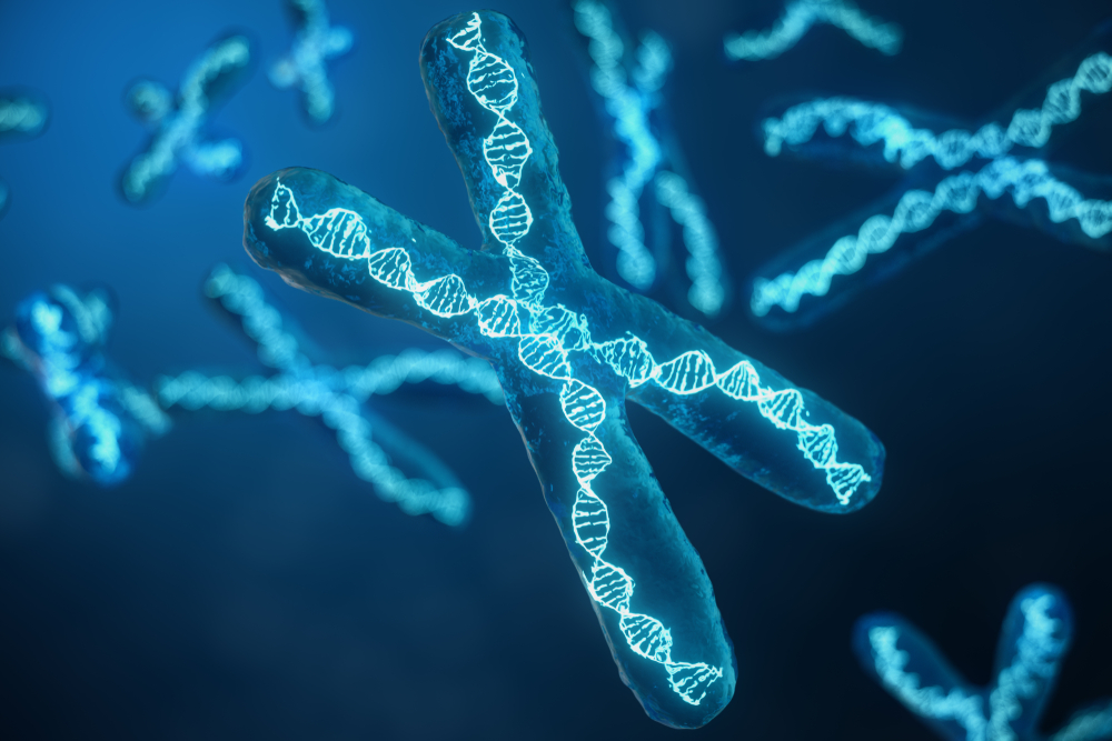
Chromosome studies look at the number and structure of chromosomes in cells. Chromosomes contain genetic information in the form of tightly packaged DNA and at certain times when cells are dividing they can be viewed under a microscope.
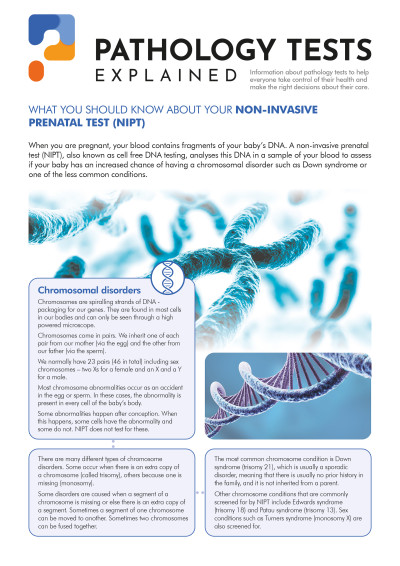
Chromosomes are normally present in 23 pairs or 46 chromosomes. One copy of each chromosome comes from our mother in the egg and the other copy from our father in the sperm. There are 22 pairs of chromosomes which are the same in both males and females with the other pair being the sex chromosomes - a pair of X chromosomes in females and one X and one Y in males.
The test can tell if the right number of chromosomes are present or if there are whole chromosomes missing or extra ones present. Sometimes, only small fragments of a chromosome are missing or duplicated. In each case the changes result in too little or too much genetic information causing health and development problems.
Large changes such as missing or extra whole chromosomes or large pieces lost or duplicated can be detected by looking at the chromosomes through a microscope. Special stains make the chromosomes appear to have bands that allows the specialist scientist to identify when pieces may be lost, gained, or rearranged and to report the changes using the bands as markers.
Very small changes may still cause problems but cannot be detected visually, in this case the studies will require a Chromosomal Microarray test which can pick up these tiny changes.
What is being tested
Chromosome studies or karyotyping is a test that evaluates the number and structure of a person's chromosomes in order to detect abnormalities. Chromosomes are thread-like structures within each cell nucleus and contain the body's genetic blueprint. Each chromosome contains thousands of genes in specific locations. These genes are responsible for a person’s inherited physical characteristics and they have a profound impact on growth, development, and function.
Humans have 46 chromosomes, present as 23 pairs. Twenty-two pairs are found in both genders (autosomes), and one pair (sex chromosomes) is present as either XY (in males) or XX (in females). Normally, all cells in the body that have a nucleus will contain a complete set of the same 46 chromosomes, except for the reproductive cells (eggs and sperm), which contain a half set of 23. This half set is the genetic contribution that will be passed on to a child. At conception, half sets from each parent combine to form a new set of 46 chromosomes in the developing foetus.
Chromosomal abnormalities include both numerical and structural changes. For numerical changes, anything other than a complete set of 46 chromosomes represents a change in the amount of genetic material present and can cause health and development problems. For structural changes, the significance of the problems and their severity depends upon the chromosome that is altered. The type and degree of the problem may vary from person to person, even when the same chromosome abnormality is present.
A karyotype examines a person's chromosomes to determine if the right number is present and to determine if each chromosome appears normal. It requires experience and expertise to perform properly and to interpret the results. While theoretically almost any cells could be used to perform testing, in practice it is usually performed on a chorionic villus sample or amniotic fluid to evaluate a fetus and on lymphocytes (a white blood cell) from a blood sample to test at other ages. Alternatively, white blood cells may be obtained from bone marrow aspirations to look for changes in individuals suspected of having haematologic or lymphoid diseases (e.g., leukaemia, lymphoma, myeloma, refractory anaemia).
The test is performed by:
 Normal male karyotype.
Example of a peripheral blood karyotype, normal male.
 Abnormal Karyotype
Example of an abnormal bone marrow karyotype showing chromosome 9 and chromosome 22 (9;22) rearrangement, indicative of chronic myelogenous leukemia (CML) or a subtype of acute lymphoblastic leukaemia (ALL)
 Trisomy 21
Example of an abnormal karyotype showing an extra chromosome 21 (Trisomy 21) indicative of Down syndrome.
Images courtesy of: Mary Lowery Nordberg, PhD AACC.
Some chromosomal disorders that may be detected include:
How is the test used?
A chromosomal karyotype is used to detect chromosome abnormalities and is therefore used to diagnose genetic diseases, some birth defects, and certain haematologic and lymphoid disorders.
It may be performed for:
a) If one or more of a woman's pregnancy screening tests, such as the first trimester Down syndrome screen or the triple or quad screen, are abnormal.b) If a pregnant woman is having amniotic fluid analysis performed because she is considered at a higher than normal risk of having a baby with a birth defect.
c) If fetal structural and/or developmental abnormalities are detected on an ultrasound.
d) If there is a known chromosomal abnormality in the family line.
What does the result mean?
Interpretation of test results must be done by a person with specialised training in cytogenetics. Some findings are relatively straightforward, such as an extra chromosome 21 (Trisomy 21) indicating Down syndrome, but others may be very complex.
Although there will be typical signs with specific chromosomal abnormalities, the effects and the severity may vary from person to person and often cannot be reliably predicted.
Some examples of abnormalities that chromosome studies may reveal include:
| Trisomy | This is the presence of an extra chromosome, a third instead of a pair. Diseases associated with trisomies include Down syndrome (associated with a Trisomy of chromosome 21), Patau syndrome (Trisomy 13), Edward syndrome (Trisomy 18), and Klinefelter syndrome (a male with an extra X chromosome – XXY instead of XY). |
| Monosomy | This is the absence of one of the chromosomes. An example of monosomy is Turner syndrome (a female with a single X chromosome – X instead of XX). Most other monosomies are not compatible with life. |
| Deletions | These are missing pieces of chromosomes and/or genetic material. Some may be small and difficult to be detected. |
| Duplications | These represent extra genetic material and may be present on any chromosome, such as the presence of two horizontal bands at a specific location instead of one. |
| Translocations | With translocations, pieces of chromosomes break off and reattach to another chromosome. If it is a one-to-one switch and all of the genetic material is present (but in the wrong place), it is said to be a balanced translocation. If it is not, then it is called an unbalanced translocation. |
| Genetic Rearrangement | In this situation, genetic material is present on a chromosome but not in its usual location. A person could have both a rearrangement and a duplication or deletion. An almost infinite number of rearrangements are possible. Interpreting the effects of the changes can be challenging. |
Duplications, deletions, translocations, and genetic rearrangements can cause a myriad of health and development issues. It depends upon what genes are missing or present in too many copies.
Some genetic rearrangements will be variations that do not cause noticeable symptoms. Balanced translocations (where two chromosomes have swapped portions of themselves but all of the genetic material is present) may cause no problems for the person who has them, but may cause problems in their children.
Many haematologic and lymphoid malignancies (e.g., leukaemia, lymphoma, myeloma, myelodysplasia) are associated with chromosomal abnormalities, which can help diagnose the disease and/or predict the clinical course of the disease.
Is there anything I should know?
Since the sex chromosomes (XX or XY) are identified during the chromosome study, this test will also, as a result, definitively determine the sex of a fetus.
Some chromosome alterations are too small or subtle to detect with karyotyping. Other testing techniques, such as fluorescent in situ hybridisation (FISH) or microarray, may sometimes be performed to further investigate chromosomal abnormalities.
It is possible for people to have cells in their body with differing genetic material. This happens because of changes early in the development of a fetus, that lead to the development of distinctly different cell lines and is called mosaicism. For example, in some cases of Down syndrome the affected person can have some cells with an extra third chromosome 21, and some cells with the normal pair.
Common questions
Chromosome studies are frequently performed, but it is not indicated as a general screening test. The majority of people will never need to have one done.
No, it requires specialised equipment to perform and expertise to interpret. In most cases, samples will be sent to a reference laboratory.
The cells that are tested must be cultured and cell division promoted. The amount of time that this takes will vary from sample to sample. Highly complex, abnormal karyotypes may require a longer time to evaluate.
More information
What is Pathology Tests Explained?
Pathology Tests Explained (PTEx) is a not-for profit group managed by a consortium of Australasian medical and scientific organisations.
With up-to-date, evidence-based information about pathology tests it is a leading trusted source for consumers.
Information is prepared and reviewed by practising pathologists and scientists and is entirely free of any commercial influence.