
Summary
What is iron and why do we need it?
Your body needs iron for daily activities, growth and development. Most importantly, it uses iron to make haemoglobin in red blood cells.
Red blood cells carry oxygen from the lungs through the bloodstream and release it to where it is needed in the body. They can do this because the iron in haemoglobin attaches to the oxygen molecules.
Our bodies cannot make iron. Iron must be absorbed from our diet or from supplements. The amount of iron absorbed is decided by our body's needs. We normally use just a little bit of iron each day.
We do not have a way of getting rid of excess iron. Instead, the body closely controls iron levels by managing how much iron we absorb.
In hereditary haemochromatosis, there is a breakdown in the process that controls iron levels, and your body behaves as if there is an iron shortage. It responds by absorbing more iron from food. Without a way to remove excess iron, it builds up in organs and tissues where over time it causes damage.
How is iron absorbed and controlled?

How the HFE gene controls iron levels
The HFE gene controls iron levels by making a protein that is also called HFE. The HFE protein controls iron absorption to keep levels in balance.
HFE protein tells the liver to make hepcidin, which is known as the master regulator of iron levels. Hepcidin controls how much iron is absorbed from your intestines and how much is released from storage.
Normally, when iron levels are high, your liver responds by making more hepcidin and releasing it into the blood. Hepcidin reduces iron absorption in your intestines and stops stored iron from being released into your blood. This lowers the level of iron in the blood.
HFE protein also stops cells from taking up transferrin and iron from the blood.
In haemochromatosis, the mutated HFE gene makes faulty HFE protein that does not function properly. This means:
Why get tested?
If you have symptoms that suggest you may have iron overload your doctor will request a Full Blood Count (FBC), which gives information on the amount of haemoglobin in your blood, as well as the size and shape of the red blood cells.
They will also request iron studies, a group of tests that looks at the iron levels in your body. This panel of tests includes ferritin, transferrin, iron and transferrin saturation.
If iron studies test results show that you have high transferrin saturation (more than 45 per cent) and a high ferritin level (more than 300 ug/L) you may have haemochromatosis. However, these are not specific for haemochromatosis and could be due to other causes. This is why genetic testing is needed to confirm the diagnosis.
Your doctor may order an HFE genetic test which can show whether there is a mutation in the HFE gene.
An inherited genetic mutation can increase the risk of developing iron overload. However, many people have these mutations but do not develop iron overload. This means the HFE genetic test cannot be used by itself to diagnose hemochromatosis, and your doctor needs to consider the results of the HFE genetic test together with your blood iron studies, liver function test results, possibly imaging of the liver and symptoms.
How HFE gene mutations are inherited
Hereditary haemochromatosis is known as an autosomal recessive disorder which means you need to inherit one mutated gene from each of your parents. It is common in people whose ancestors came from northern Europe. Studies have shown that up to half of people who inherited two mutated copies (C282Y) of the HFE gene develop iron overload and have test results that show high levels of ferritin and transferrin saturation. But only about 10 - 30 percent of these people develop symptoms of haemochromatosis such as fatigue, joint pain and organ damage.
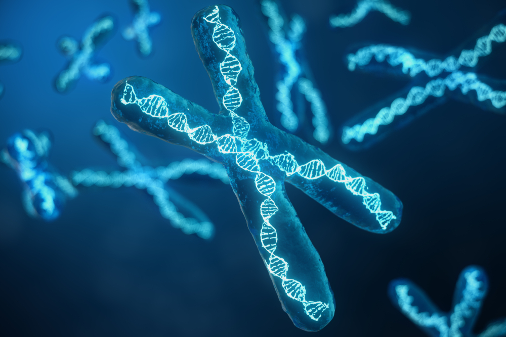
Our bodies are made up of billions of cells. Almost every cell has a nucleus (a sac in the middle of the cell) containing a complete set of genetic material. Genes give the instructions to make proteins that tell our bodies how to function.
Genes are made from a substance called DNA. Inside the nucleus, strands of DNA are wound up tightly into structures called chromosomes.

We inherit one set of 23 chromosomes from our mother (via the egg), and another set of 23 chromosomes from our father (via the sperm).
Sperm and egg cells are different from normal cells. They have only 23 chromosomes, which is half the number of chromosomes in normal body cells. So, when a sperm cell and an egg cell fuse in conception, they each bring together 23 chromosomes to form a fertilised egg that has a full set of 46 chromosomes. Combining genetic information from both parents in this way ensures genetic diversity but may also result in the mutated gene being passed on to your children.

Having the test
Sample
Blood.
Any preparation?
None.
Your results
In order to study them, scientists have numbered chromosome pairs from 1 to 22, with the 23rd pair being the sex chromosomes labelled as XX or XY. The HFE gene is on chromosome 6.
Mutations are named according to their location on the gene.
C282Y
The most common mutation that causes haemochromatosis is C282Y. Most cases of haemochromatosis occur in people who have two copies of the C282Y mutation, one on each of the pair of chromosomes. If you inherit C282Y mutations in both copies of the HFE gene, you are at risk of developing haemochromatosis, although many people do not develop the disease.
H63D
Another mutation, H63D is associated with a more moderate risk of developing haemochromatosis. People who have two copies of the H63D mutation may have a very slightly increased risk of developing haemochromatosis, while people who have only one copy of any of these mutations and one normal gene are not at increased risk.
S65C
A third mutation, S65C may lead to mild to moderate iron overload but this does not cause haemochromatosis.
Compound heterozygotes
Occasionally, haemochromatosis occurs in people who have one abnormal copy of C282Y and one of either H63D or S65Y. These are known as compound heterozygotes and can cause mild or moderate iron overload. Level of iron overload in these cases usually depends on other factors such as alcohol use or the presence of other liver disease.
S65C is rarely tested for but when it occurs in combination with C282Y may cause iron overload in some people. The S65C mutation by itself will not cause raised levels of transferrin saturation or ferritin.
Single mutation
Some people with a single mutation for C282Y or H63D have high transferrin saturation and ferritin levels but do not develop the complications related to iron overload.

| The range of results of the HFE genetic test and how they relate to the level of risk of developing haemochromatosis. | ||
| Result | Genes | Risk of developing haemochromatosis |
| Two copies of the C282Y mutation | C282Y/C282Y | Highest risk |
| One copy of C282Y mutation and one copy of H63D mutation | C282Y/H63D | Increased risk |
| Two copies of the H63D mutation | H63D/H63D | Slightly increased risk |
| One copy of C282Y mutation and one copy of S65C mutation | C282Y/S65C | Slightly increased risk |
| One copy of C282Y/H63D/S65C mutation and one normal gene | Carrier state (heterozygous) | No increased risk |
Secondary haemochromatosis
Other conditions can cause iron overload. The most common causes are liver disease, thalassaemia and blood transfusion-related iron overload.
Further testing
An MRI liver scan or a liver biopsy may sometimes be required to diagnose iron overload and provide further information. These tests are used to assess the hepatic iron index (the amount of iron in the liver) and look for fibrosis and cirrhosis.
Family testing
First-degree relatives such as parents, brothers, sisters and children of people with known haemochromatosis are at high risk of developing the disorder and should be offered an iron studies test and an HFE gene test, even if they do not yet have high ferritin levels. If a relative has a genetic makeup that suggests they are at risk of developing haemochromatosis, they can be closely monitored and started on treatment early in the disease process, if it is required.
More to know?
About 100 mutations in the HFE gene have been recognised but most of these are very rare and the clinical significance of many of them is not yet known. Only C282Y and H63D mutations are commonly tested for. In some circumstances, your doctor may request that the laboratory looks for rare mutations.
Mutations in other genes may cause haemochromatosis. These are
Haemochromatosis caused by mutations in these genes is very rare and genetic testing is not routinely available and does not attract a Medicare rebate. Referral to a specialised haemochromatosis clinic or clinical genetics service is recommended.
Treatment of haemochromatosis is usually straightforward. It involves the regular removal of blood known as a venesection, which is similar to a blood donation. This removes excess iron from the body.
Questions to ask your doctor
The choice of tests your doctor makes will be based on your medical history and symptoms. It is important that you tell them everything you think might help.
You play a central role in making sure your test results are accurate. Do everything you can to make sure the information you provide is correct and follow instructions closely.
Talk to your doctor about any medications you are taking. Find out if you need to fast or stop any particular foods or supplements. These may affect your results. Ask:
More information
Pathology and diagnostic imaging reports can be added to your My Health Record. You and your healthcare provider can now access your results whenever and wherever needed.
Get further trustworthy health information and advice from healthdirect.
What is Pathology Tests Explained?
Pathology Tests Explained (PTEx) is a not-for profit group managed by a consortium of Australasian medical and scientific organisations.
With up-to-date, evidence-based information about pathology tests it is a leading trusted source for consumers.
Information is prepared and reviewed by practising pathologists and scientists and is entirely free of any commercial influence.